Our Science

Reimagining Treatments for Vision Loss and Beyond.

Regenerative medicine in the development of novel treatments for diseases focuses on methods to regrow, repair, or replace damaged or diseased cells, tissues, or organs. The approaches to develop new regenerative medicine treatments vary widely from the use of materials or de-novo generated cells in tissue engineering, to cell therapies such as the activation of progenitor cells, to immune-modulation and gene-editing and gene-therapy based approaches to induce tissue repair and or new cellular development. Regardless of the method undertaken, the ultimate goal of regenerative medicine is the same; to repair or replace tissue or organ function lost due to disease, damage or age, in both rare and common diseases. At Caeregen Therapeutics, our science focuses on the basic and systems biology related to the target tissues and organs of interest, the microenvironments comprising these structures, and the restoration of normal cellular signaling or processes that to healthy tissue and organ function.

APPROACH

Vision loss due to retinal damage is an urgent, global, unmet medical need...

DISCOVERY PROCESS

Norrin participates in chemical signaling pathways that affect the way cells and tissues develop...

INHERITED DISEASES

FEVR is a life-long hereditary retinal vascular disease, which occurs as a consequence of a biochemical imbalance between vascular endothelial growth factor and tissue growth factor beta...

ACQUIRED DISEASES

Retinal vein occlusions are small “strokes” in the retina caused by blockages in the veins draining the retinal circulation...

APPROACH

Vision loss due to retinal damage is an urgent, global, unmet medical need. Damage to retinal capillaries and neuronal tissue is a leading cause of vision loss and impairment. An epidemic looms with a large and growing aging population and continuing increases in chronic illnesses like cardiovascular disease, diabetes, and obesity, all conditions that often lead to retinal damage. We characterize retinal damage and vision loss induced by such conditions as an ‘acquired’ progressive disease. The range of inherited diseases associated with retinal-related vision loss are described in the “Inherited Diseases’ section here.

Current treatments only slow progression while patients are receiving them. Retinal disease involving capillary leakage or dropout has been treated by laser and surgical therapy in the past, to stabilize the retinal damage. In 2004, a class of drugs targeting vascular endothelial growth factor, or VEGF, was introduced for neovascular age-related macular degeneration (NVAMD), and subsequently macular edema due to retinal vein occlusion (RVO) and diabetic retinopathy (DME). When vessels are damaged, they signal the body to regrow using VEGF, resulting in prolific overgrowth that causes further damage. Anti-VEGF drugs have been used to reduce the abnormal “leakiness” of blood vessels in eyes with retinal disease. These drugs were approved by the FDA for use in NVAMD, RVO and DME, but also have been used in an off-label fashion in people affected with other progressive retinal diseases. The result is an inhibition of abnormal vascular growth and reduction in leakage from damaged retinal capillaries which slows degeneration of retinal tissue. However, Anti-VEGF therapy impedes retinal capillary regeneration. This loss of tiny retinal blood vessel—capillary—function which results in vision loss as retinal sells essential for vision are deprived of oxygen and nutrients essential for normal function. The primary limitation of both approved and investigational anti-VEGF therapies is that they do not stimulate capillary regrowth. New therapies are urgently needed that repair cellular junctions and transmembrane leakage, regenerate neuronal and capillary cells, and protect against capillary and neuronal damage and loss. This challenge has led the Caeregen Therapeutics team to develop its first drug therapy, Noregen®. Noregen® is a synthetic protein modeled after the naturally occurring protein norrin that guides retinal vascular development in the earliest stages of life.

Pipelines & Programs

Caeregen Therapeutics is developing a revolutionary approach to reverse capillary and neuron loss in both inherited and acquired retinal disease.

Discovery process

Norrin participates in chemical signaling pathways that affect the way cells and tissues develop. Studies show that norrin plays a role in Wnt signaling, which is important for cell division (proliferation), attachment of cells to one another (adhesion), cell movement (migration), and many other cellular activities.


Norrin is one of many proteins, or ligands, that can attach (bind) to other proteins called frizzled receptors. These receptors are embedded in the outer membranes of cells. Norrin binds with the receptor frizzled-4 (produced from the FZD4 gene), fitting together like a key in a lock. When a molecule like norrin binds to the frizzled-4 receptor (molecules that bind to receptors are called “ligands”, the binding of ligand to a receptor initiates a series of chemical steps termed signal transduction or “signaling”, and frizzled-4 belongs to a family of receptors known as the “Wnt signaling family” which are critical for blood vessel development), it initiates a multi-step process that regulates the activity of certain genes.


The norrin protein and its receptor frizzled-4 participate in developmental processes that are believed to be crucial for normal development of the eye and other body systems. Although Wnt signaling occurs in all cells of the body, loss of norrin cannot be replaced by other Wnt ligands. In particular, norrin seems to play critical roles in the specialization of cells in the retina (the thin layer at the back of the eye that senses light and color) and the establishment of a blood supply to the retina and the inner ear.


Norrin binds the Wnt receptor frizzled-4 and induces pathway activation in conjunction with cell surface co-receptor LRP5. This ligand is known to be involved in angiogenesis since overexpression in norrin-deficient mice induces the growth of ocular capillaries and inactivating mutations results in Norrie disease which is characterized by ocular vascular defects, impaired cochlear vascularization, and central nervous system abnormalities. Abnormalities in Fz4 or LRP5 result in defects in retinal vascular development and in a phenotypically similar condition, familial exudative vitreoretinopathy (FEVR). A third component that inhances norrin signaling in blood vessels is TSPAN12 molecule. TSPAN12 acts as a chaperone in the norrin receptor complex thereby signaling norrin through nuclear beta-catenin, through a new Wnt driven signaling cascade.

Norrin activated retinal progenitor cells (green) integrate into injured retina
Macular involving tractional retinal detachment (Stage 4 FEVR) Caption for B&W photo: Flourecene angiogram demonstrating a vascular peipheral retina (Stage 1 FEVR)

Inherited diseases

Familial Exudative Vitreoretinopathy (FEVR)

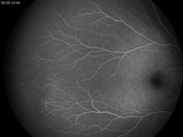
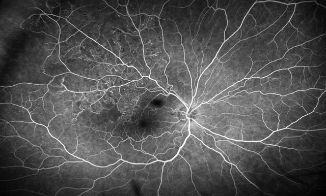
FEVR is a life-long hereditary retinal vascular disease. Impaired angiogenesis in the retina of young children can lead to aberrant neovascularization and symptoms, such as the formation of retinal folds and tears, retinal detachment, or even total vision loss.1 Among the genes associated with FEVR, components of the Wnt/β-catenin signaling pathway are highly prevalent. These are the genes encoding the growth factor Norrin (NDP), the receptor Frizzled-4 (FZD4), the low-density lipoprotein receptor-related protein (LRP5), and tetraspanin-12 (TSPAN12), which forms transmembrane receptorcomplexes with LRP5 and Frizzled-4.


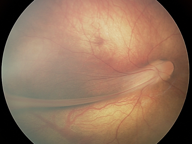
Familial exudative vitreoretinopathy (FEVR) was first described by Criswick and Schepens.It is a rare disorder of retinal blood vessel development principally affecting retinal angiogenesis, leading to incomplete vascularization of the peripheral retina and poor vascular differentiation. Cases can be inherited in an autosomal dominant, autosomal recessive, or X-linked manner, or can affect individuals with no family history. Most FEVR patients have an avascular peripheral retina but expressivity may be asymmetric and is highly variable, ranging from asymptomatic to severe within the same family. If there is a significant degree of retinal ischemia, then secondary neovascularization can occur leading to fibrosis, traction of the posterior pole structures, retinal detachment (RD), retinal folds, or complete retinal dysplasia in the most severe cases. A diagnosis of FEVR can be made if there is evidence of peripheral retinal avascularity in Z1 eye in patients of any age who were born at full term, or preterm with a disease tempo not consistent with retinopathy of prematurity (ROP).


Five genes have so far been identified that when mutated, cause FEVR; NDP (MIM 300658), FZD4 (MIM 604579), LRP5 (MIM 603506), TSPAN12 (MIM 613138), and ZNF408. Mutations in these genes account for around 50% of FEVR cases.3 Four of the genes identified to date have been shown to have a role in a variant of the Wnt signaling pathway, termed as Norrin/Frizzled4 signaling suggesting a crucial role for this pathway in retinal vascular development.


Currently however, there are no approved pharmacological therapies for FEVR.


1 Park, H., Yamamoto, H., Mohn, L. et al. Integrin-linked kinase controls retinal angiogenesis and is linked to Wnt signaling and exudative vitreoretinopathy. Nat Commun10, 5243 (2019). https://doi.org/10.1038/s41467-019-13220-3

2, 3 Gilmour, D. Familial exudative vitreoretinopathy and related retinopathies. Eye 29, 1–14 (2015). https://doi.org/10.1038/eye.2014.70

Acquired diseases

Retinal Vein Occlusion (RVO)

Retinal vein occlusions are blockages in the vessles draining the retinal circulation. There are two types: central retinal vein occlusions (CRVO) and branch retinal vein occlusions (BRVO) depending on larger or smaller vessels in the retina. In many cases, the cause is from hardening of the arteries, which run adjacent to the veins and compress them akin to stepping on a garden hose. This blocks blood drainage from the retina and can cause ischemia (lack of blood flow) hemorrhages and fluid leakage (macular edema) in the retina which can be visually debilitating. Patients may have risk factors including uncontrolled high blood pressure, obesity, heart disease, or glaucoma. Some younger patients may have blood clotting disorders.

RELEVANT PUBLICATIONS